Research
Electrosynthesis with BDD Electrodes to Produce Value-Added Products.
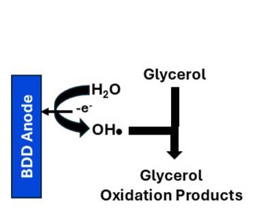
Boron-doped diamond (BDD) thin-film electrodes are next-generation sp3-bonded carbon materials. They exhibit excellent electrochemical properties, have high chemical stability, and are inexpensive and available commercially making them suitable for sensing, water treatment, energy conversion, and electrosynthesis. Electrosynthesis is a process in which electrical energy is used to drive oxidation or reduction reactions enabling the efficient and sustainable production of value-added products. Electrosynthesis can replace more traditional and polluting processes and can be conducted under mild conditions. BDD electrodes have two properties critical for electrosynthesis: (i) dimensional stability and degradation resistance and (ii) a wide working potential window in aqueous electrolyte solutions; a property that enables electrochemical reactions to be performed over a wider potential range than is possible with other electrodes (i.e., less interference by deleterious solvent-electrolyte electrolysis). In this project, fundamental research will be conducted to demonstrate that BDD electrodes can function efficiently and stably for the electrooxidation of glycerol to produce value-added products. Glycerol is primarily produced as an abundant, low-cost by-product of biodiesel manufacturing. Due to a global surplus, there is a technological need to develop a sustainable method to convert crude glycerol into a variety of high-value chemicals.
The electrooxidation of glycerol can yield C1, C2, and C3 organic compounds, with product selectivity that depends on the BDD electrode material properties and reaction conditions (potential, electrolyte composition and pH). A range of value-added products can be produced, including dihydroxyacetone (DHA), glyceric acid (GLA), formic acid (FA), lactic acid, glycolaldehyde, and oxalic acid. These products have value in diverse applications across multiple industries. Research is being conducted to better understand how the electrode microstructure, boron-doping level, surface chemistry, applied potential and current density, solution conditions (aqueous electrolyte and pH), temperature, and flow affect the oxidation of biomass-derived glycerol to form different products.BDD Glycerol OxidationGlycerol Oxidation Products
Towards an Electrochemical Immunosensing Platform for Monitoring Biomarkers of Lung Cancer and Chemotherapy-Induced Cardiac Dysfunction in Exhaled Breath Condensate Liquid Biopsy.
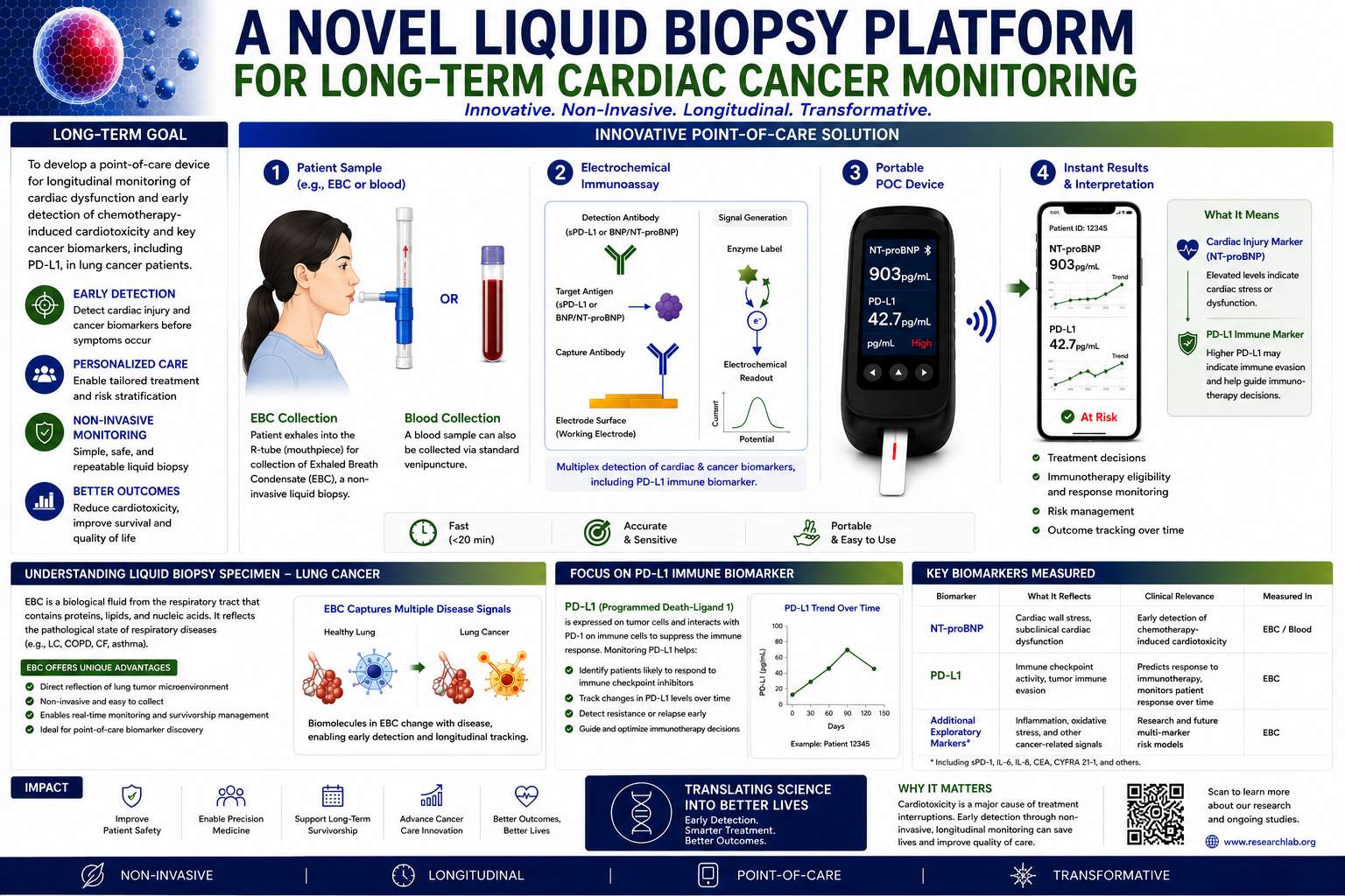
Research is being conducted on first-generation, point-of-care diagnostic technology useful for the clinical monitoring, care delivery, and disease management of human subjects with lung disease.
One project aim is to establish the clinical value of exhaled breath condensate (EBC) as a transformative, non-invasive liquid biopsy specimen for lung cancer (LC) disease management. Building on this innovative platform, we are working to prototype two first-in-class electrochemical immunosensing assays designed for longitudinal, point-of-care monitoring of critical biomarkers.
The first sensing assay targets soluble PD-L1 (programmed death ligand 1), the separated fragment of an immune checkpoint protein overly expressed by tumor cells that is a validated predictor of response to immune checkpoint inhibitor therapy, particularly in non–small cell lung cancer (NSCLC). In other words, sPD-L1 is a biomarker of NSCLC. By enabling serial sPD-L1 measurements in real time, this technology has the potential to overcome current limitations of tissue-based assays, offering a dynamic and personalized approach to immunotherapy management and assessing the effectiveness of treatment on the cancer.
Hypothesis 1: sPD-L1 levels in EBC will be significantly higher in LC patients at diagnosis than in healthy controls and will decrease over time in patients with effective treatment.
The second assay measures BNP (β-type natriuretic peptide), a sensitive biomarker of early cardiac injury and a strong predictor of chemotherapy-induced cardiotoxicity. Some chemotherapy drugs used in cancer treatment can cause cardiac damage and BNP is an indicator of this damage. Early identification of subclinical cardiac damage could allow timely intervention, reduce treatment-related morbidity and enable safer delivery of life-prolonging therapies.
Hypothesis 2: BNP levels in EBC will be significantly higher in LC patients experiencing chemotherapy related cardiac dysfunction than in healthy controls.
Despite recent advances in LC therapeutics, current diagnostic and monitoring approaches remain constrained by invasiveness, cost, and limited ability to provide real time longitudinal insight. By targeting both tumor immune biology (sPD-L1) and treatment related cardiotoxicity (BNP), this project unifies precision oncology and cardio oncology within a single, scalable monitoring framework.
If successful, this work will deliver novel diagnostic tools that integrate seamlessly into clinical workflows, advancing precision oncology and survivorship care. Ultimately, these innovations have the potential to redefine biomarker monitoring by shifting from invasive, static assessments to non-invasive, dynamic, and patient-friendly diagnostics, thereby improving outcomes for patients with lung cancer.
Chemically Modified Carbon Surfaces - Impacts on Electrochemical Reaction Kinetics.
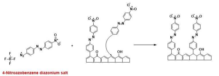
The surface chemistry of carbon materials, such as diamond, glassy carbon, carbon nanotubes and graphene, and metals (Au, Pt, Ni) can be controlled by bonding organic layers, such as through the use of aryl diazonium salts. Reductive electron transfer to the diazonium molecule in solution results in the homolytic cleavage of dinitrogen and the generation of an aryl radical. The radical formed at the electrode-solution interface subsequently bonds to the surface through carbon-carbon and metal-carbon bonds. Aryl diazonium adlayers can be formed in aqueous and non-aqueous solution using electrochemically-assisted and spontaneous formation processes. The spontaneous formation involves simply immersing the substrate in a diazonium solution under open circuit conditions for a fixed time. The attachment of 4-nitroazobenzene diazonium is shown in this example.
A variety of sp2 and sp3 carbon electrodes have been modified using the electrochemically assisted formation of the adlayer. Much less studied is the spontaneous formation of the aryl diazonium adlayers without electrical bias under open circuit conditions. The spontaneous formation of aryl diazonium adlayers on carbon electrodes is important because it provides a simple, versatile, and potentially scalable route for modifying such surfaces without the need for applying an electrochemical potential. These controlled surface modifications have implications for electrochemical sensing, corrosion protection, and biomedical interfaces. However, many fundamental questions remain regarding the mechanism, structure, and stability of the resulting films. This innovative and impactful research project will address this knowledge gap.
This research is investigating the role of carbon electrode microstructure (sp2 pyrolyzed phororesist thin films (PPF) and sp3 boron-doped nanocrystalline diamond thin films), aryl diazonium molecule type, immersion time, aryl diazonium molecule solution concentration and solvent (aprotic CH3CN and protic H2SO4) on the adlayer formation kinetics, adlayer coverage, adlayer stability, and impact on electrochemical reaction kinetics (i.e., catalyzing or blocking). A comprehensive approach will be used to understand the fundamental aspects of spontaneous adlayer formation. The adlayer formation kinetics and adlayer coverage as a function of aryl diazonium molecule type, immersion time, aryl diazonium molecule solution concentration and solvent will be characterized using confocal Raman microprobe spectroscopy, x-ray photoelectron spectroscopy (XPS) static contact angle measurements and long-optical pathlength spectroelectrochemistry. The adlayer morphology and defect density will be probed using ex situ and electrochemical tapping mode AFM and scanning Kelvin probe microscopy (SKPM). Electrochemical reactions occurring through defects in the adlayer or through the adlayer itself will be studied spatially using scanning electrochemical microscopy (SECM). The promoting or blocking effects of the adlayers will be assessed using multiple surface sensitive and insensitive redox and a suite of electrochemical measurements including rotating disk voltammetry. These effects will be quantitatively evaluated from the electron-transfer and mass-transfer kinetics. The intellectual merit of a comprehensive study of the spontaneous aryl diazonium adlayer formation process lies in its potential to advance fundamental understanding of interfacial electron transfer, surface reactivity, and covalent surface modification on carbon materials.
Understanding the Effects of Micro and Nanoplastic Particles on Neurosignaling and Function in the GI Tract.
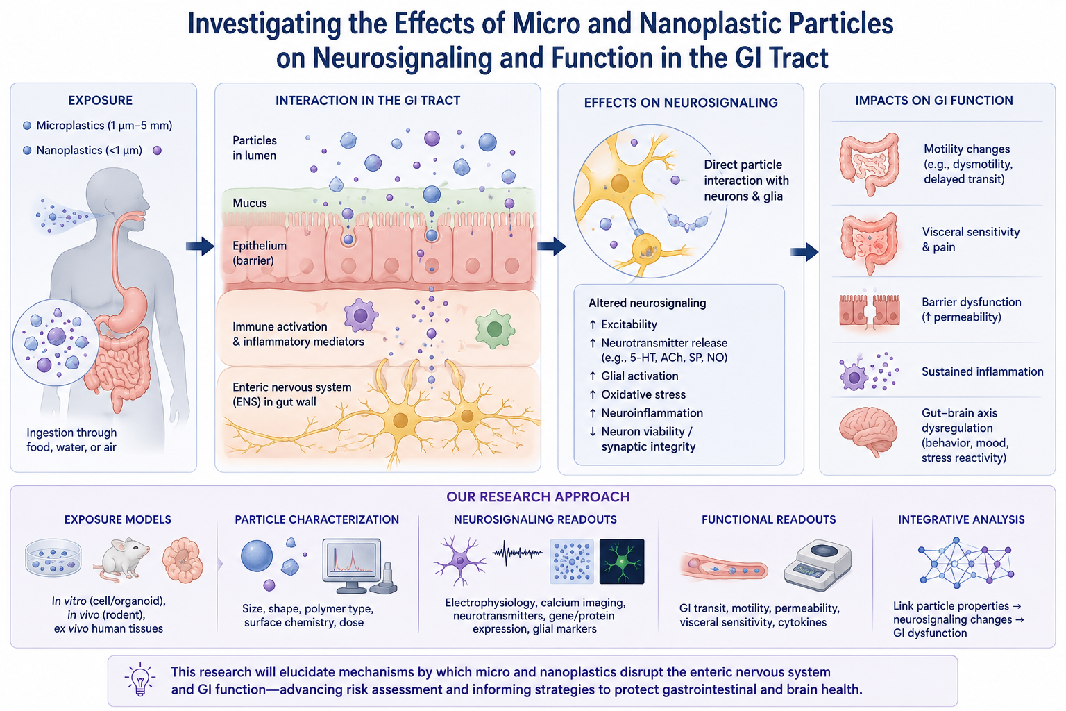
Microplastics are particles, films, and fibers with diameters from 1 µm up to 5 mm. Microplastics get broken down in the environment into nanoplastic particles (below 1 µm) via weathering processes. Nanoplastic particles are also used in various commercial products. Given their increasing prevalence in the environment and the heightened concern over the health risks to humans, we are proposing some innovative and timely research to better understand how chronic exposure impacts neurosignaling processes and function in the gut. These plastic particles enter the body via ingestion through the food we eat and the water we drink. Therefore, their toxicological effects are expected to manifest themselves initially in terms of alterations in neurogastrointestinal signaling and function. Researchers have discovered microplastics present in the blood of patients, embedded deep in lung tissue, and in placentas. Once inside the body, these plastic particles can accumulate in various organs, tissues, or the digestive system. The full extent of their impact on human health is still unknown, but potential concerns include toxicity, inflammation, and disruption of biological processes. Nanoplastic particles (< 1 µm) are an even greater concern because of their ability to easily penetrate cells and tissues more so than larger microplastics, potentially leading to more significant biological impacts. Smaller nanoplastic particles tend to show greater biological reactivity than larger microplastics, yet toxicity likely depends strongly on context (material, dose, chemistry, and exposure route).
There is a knowledge gap regarding how micro and nanoplastic particles are transported and accumulated in the gut wall and what their impact is on neurogenic signaling and how the GI tract processes food, absorbs nutrients, and eliminates waste. Proper neurogenic signaling is crucial for regulating gut motility, secretion, and overall digestive function.
Hypothesis: It is hypothesized that these plastic particles will accumulate in a dependent manner (material type, particle size and dose) in nerve and secretory cells over time and that chronic exposure will cause dysfunction in neurogenic signaling pathways through oxidative stress/inflammation mechanisms leading to abnormal gut function. Such dysfunction may lead to digestive disease.
In a project soon to be started, our experienced interdisciplinary team will seek to initiate a new multi-investigator project on this priority topic. We will investigate the biological effects of chronic exposure to micro (1 µm diam.) and nanoplastic (100 and 10 nm diam.) particles in drinking water applied ad libitum to both male and female guinea pigs. Two plastic materials will be studied: polystyrene and polycarbonate. Experiments will begin on animals at 6 weeks of age as they possess a mature enteric nervous system at this age. A particle concentration of 100 µg/mL in drinking water will be used. Measuring the volume consumed will enable us to determine the dose of particles consumed by each animal. The study will be designed to (i) understand the distribution particles in the small and large intestine and their interaction with epithelial, immune and enteric nerve cells, (ii) learn the effects on neurotransmission (serotonergic, nitrergic, purinergic and cholinergic) and enteric neuron excitability, synaptic function and glial cell activation, and (iii) assess the impacts on altered motility, inflammation/oxidative stress and barrier dysfunction.
Overall, the research will elucidate how micro and nanoplastic particles disrupt enteric neurotransmission and GI function providing critical insights on risk assessment and strategies for protecting gastrointestinal health. This innovative and potentially impactful research project will involve three broad specific aims. Animals receiving plastic particle-free drinking water will be used as controls.
Development and Physiological Testing of ATP Biosensors for Assessing Kidney Physiologic Function.
ATP is a molecule that provides energy for a plethora of enzymatic reactions in the cell. It is also an important humoral factor responsible for para- and autocrine signaling. In the kidney, ATP plays a major role in orchestrating renal autoregulation, hemodynamics and epithelial transport. ATP functions as both an excitatory and inhibitory neurosignaling molecule in the GI tract regulating multiple digestive functions. Measurement of extracellular ATP dynamics in vivo is a desired analysis in different scientific fields and represents a technical advance. Dynamic ATP measurements in vivo provide insight into how cells regulate energy production and consumption in real time, which is essential for understanding metabolism, physiology, and disease processes. Electrochemical sensors and biosensors are well suited for real time measurement of ATP providing the ability to track rapid changes in concentration near sites of release and action. Real-time measurement would improve disease detection, treatment, and understanding of many physiological processes.
In this new project we will (i) develop an electrochemical ATP biosensor suitable for physiological experiments. The first generation of the biosensor that utilizes glucose oxidase (GOx) and hexokinase (Hex) immobilized within the electroactive polymer, polyethylenedioxythiophene (PEDOT), is being fabricated and tested in standard solutions to determine the ATP detection figures of merit (sensitivity, response time, reproducibility). We will then (ii) apply the ATP biosensor in vivo to confirm that blockage of pannexin-1 ion channels in the kidney precludes extracellular ATP release. This protein forms a hemichannel and is predicted to be permeable for ATP. Pannexin-1 knockout mice and unique pharmacological inhibitors are available for testing this hypothesis. This a new project being performed collaboratively with Dr. Tengis Pavlov - Henry Ford Health. Earlier publications from the Pavlov group demonstrated feasibility of electrochemical biosensors to measure ATP in the kidneys of anesthetized animals in vivo.